Spina Bifida
Clinical data
- An eighteen day old infant was admitted with a ruptured spina bifida and signs of meningitis, viz. neck rigidity and convulsions.
Post-mortem pathology
-
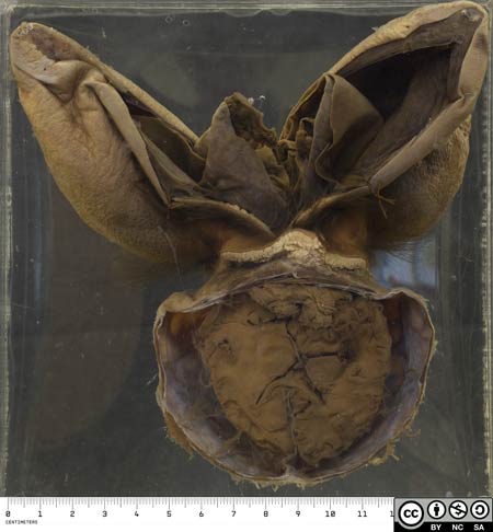
At post mortem there was a large cystic mass over the sacrum, covered in part by skin and in part by necrotic sloughing tissue. The specimen shows this lesion (excised) alongside the child’s brain.

- This is a section through the baby’s pelvis.
- Anteriorly lie the pelvic organs.
- Posteriorly, the spinal membranes and spinal cord bulge out through the unfused spinal vertebrae.

- The greatly thickened cauda equina passes out to be incorporated in the posterior wall of the cyst.
- A pyogenic membrane lines the surrounding cyst.

- A suppurative exudate is present in the subarachnoid space of the brain, especially around the midbrain and pons.

- This is a myelomeningocoele (or meningomyelocoele) since both meninges and neural tissue have extended though the defect in the spinal column.
- The major complications are neurological deficit in the lower limbs and in bowel and bladder control, as well as infection, which in this case has ascended to cause a fatal meningitis.
Related Specimens


