Digital Pathology by University of Cape Town is licensed under a Creative Commons Attribution-NonCommercial-ShareAlike 4.0 International License. Permissions beyond the scope of this license may be available at www.pathologylearningcentre.uct.ac.za.


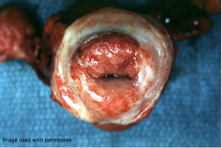
Early invasive cervical squamous carcinoma - removed by
hysterectomy
Image used with permission, Image 1350 from Peir Digital Library – public
access image database,
http://peir.path.uab.edu University of Alabama at Birmingham, Department of Pathology

Advanced cervical carcinoma with urinary obstruction - removed at
autopsy
Clinical data
-
The patient was an elderly woman who presented with post-menopausal bleeding, a foul smelling vaginal discharge, pain in the lower abdomen, dysuria and urinary frequency.
-
On vaginal examination a large fungating growth of the cervix was found.
-
She was given radiotherapy (palliative) but two weeks later she became mentally confused, then comatose, and died.
At autopsy
- The specimen shows, in the lower half, the bisected uterus and cervix (flapped open) and base of bladder.
- All these are extensively infiltrated by tumour tissue.

- Just above the bladder the right ureter disappears into tumour.
- Both ureters are dilated, the right more so.
- The right kidney is atrophied while the left kidney is enlarged.
- This indicates that obstruction of the right ureter preceded that of the left by a long enough interval to allow some compensatory hypertrophy of the left kidney.

Comment
- Most patients with stage IV carcinoma of the cervix die in uraemia (renal failure) – this is explained by the anatomical proximity of the ureters to the cervix, just before they enter the bladder.
- Advanced cervical carcinomas extend directly into contiguous tissues – vagina, rectum, bladder, ureters, paracervical tissue.
- Obstruction of the ureters by tumour extension causes back pressure on the kidney, with dilatation of the renal pelvis and calyx, and atrophy of the functional renal parenchyma (=hydronephrosis).
- Infection (pyelonephritis) often supervenes.
A useful link
- Run through images 1-15 (covering cervix and vagina) on this WebPath page http://library.med.utah.edu/WebPath/FEMHTML/FEMIDX.html#2

