Digital Pathology by University of Cape Town is licensed under a Creative Commons Attribution-NonCommercial-ShareAlike 4.0 International License. Permissions beyond the scope of this license may be available at www.pathologylearningcentre.uct.ac.za.
Clinical data
- This is an example of a traumatic vascular injury in the central nervous system.
- Unfortunately there is no clinical data for the specimen.

Post mortem pathology
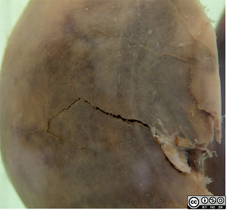
- A side view of the specimen shows a fracture in the left parietal region.
- This is a common site for a skull fracture.
- The pterion is the weakest part of the skull http://en.wikipedia.org/wiki/Pterion
- The anterior branch of the middle meningeal artery runs through the dura beneath the pterion

- The skull fracture has resulted in laceration of artery,
- followed by a massive arterial haemorrhage.
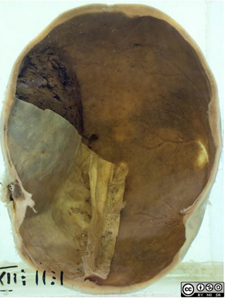
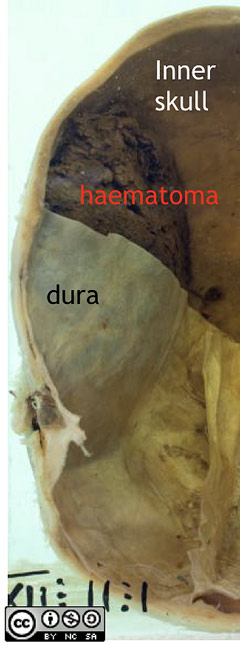
- The haematoma which formed has a smooth convex profile.
- The haematoma would have compressed the underlying brain, resulting in pressure effects leading to the patients death.

- On the opposite undamaged side of our specimen the pterion can be seen as a thin, almost transparent area of skull.
- The grooves made by branches of the middle meningeal are seen on the inner skull in the same region
- (The grooves of the anterior and posterior meningeal arteries can also be seen.)

- Note the location of the haematoma, it lies between the dura and the internal surface of the skull i.e. it is epidural or extradural .
- (Under arterial pressure the expanding haematoma stripped the dura from the periosteum.)
- Contrast this with a subdural haematoma, which would collect between the dura and the surface of the brain

Subdural haemorrhage link
- See a case of subdural haemorrhage in the collection.

