SYSTEM: CARDIOVASCULAR
FREQUENCY: 4 per 10 000 live births
PATIENT HISTORY: No clinical data were recorded
SPECIMEN C1-c24-0653
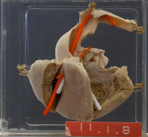
This specimen shows the four components of the tetralogy of Fallot:
- Sub-pulmonary stenosis (stenosis just below the pulmonary valve), indicated by the thin orange probe.
- Hypertrophy of the right ventricle, a consequence of the obstruction to the pulmonary outflow.
- Ventricular septal defect; the bent white probe passes from the left to right ventricle through this defect.
- Overriding aorta (the aorta lies over the ventricular septal defect and connects to both left and right ventricles),shown by the large orange probe which passes from the right ventricle into the aorta.


THE CONDITION:
In Fallot's tetralogy blood enters the aorta from both left and right ventricle, so a mix of oxygenated and unoxygenated blood is sent to the body, and the infant usually presents with cyanosis soon after birth*. Because of the obstruction to the pulmonary outflow tract, which worsens with age, there is also preferential flow into the aorta, known as right to left shunt. The four components of tetralogy of Fallot vary from patient to patient, and so the severity of the condition varies. Inevitably right heart failure develops, and ultimately biventricular failure. Without treatment the mortality is high in the first year of life and few patients will survive to adulthood (approximately 20% alive at age 10 years, 10% alive at age 20 years and 3% at 40 years).
*Other clinical findings in TOF infants are a heart murmer, difficulty in feeding and failure to thrive. In older children there will be dyspnoea on exertion, clubbing of fingers and toes and polycythaemia. They may also suffer from episodes of syncope and are at risk for sudden death. A typical behaviour in children with TOF is squatting, which increases peripheral vascular resistance and so improves pulmonary blood flow. On X-ray the TOF heart is classically boot or clog shaped, and the full anatomical picture is obtained by echocardiography.
AETIOLOGY:
TOF is seen in other mammals such as horses and rats.
Most cases of TOF are sporadic and non-familial and the aetiology is said to be "multifactorial". There is a small (≤ 5%) risk for siblings of an index case. Extra-cardiac anomalies are found in some patients. There is an association with Down's syndrome.
PREVENTION & TREATMENT:
Today, corrective cardiac surgery can be done early in life; optimally at around 12 months. Cyanosed and hypoxic babies awaiting surgery are treated with prostaglandins to keep the ductus arteriosus open, which allows additional mixing of unoxygenated and oxygenated blood. Survival and long-term outcome depend on the particular anatomical defects of the individual patient, and on the surgical expertise available. Many patients do very well and have no problems in later life other than reduced exercise tolerance. Follow-up by a cardiologist is essential; arrhythmia is an important post-surgical complication. Some patients require repeat operation for residual defects. Later problems are pulmonary incompetence and heart failure. Endocarditis is a lifelong risk requiring antibiotic prophylaxis.

|

|
| A self portrait by the Dutch artist Dick Ket (1902 - 1940) who is believed to have had Fallot's tetralogy with dextrocardia. [Dextrocardia is where the heart is in the right side of the chest with a right-sided apex; there are often associated intracardiac anomalies.] | Note the clubbing of his fingers. |
