Digital Pathology by University of Cape Town is licensed under a Creative Commons Attribution-NonCommercial-ShareAlike 4.0 International License. Permissions beyond the scope of this license may be available at www.pathologylearningcentre.uct.ac.za.
Clinical data
-
The patient was 6 years old and had just started treatment for spinal tuberculosis.
Post mortem pathology
- The specimen is the heart and lungs of a child.
- Numerous pale miliary foci, all ≤ 1mm in diameter, are uniformly distributed throughout both lungs


Some comments

Photo: Elke Wetzig
From: https://commons.wikimedia.org/wiki/File%3AHirsekoerner.jpg
- Chest x-ray in miliary tuberculosis: http://www.meddean.luc.edu/lumen/MedEd/medicine/pulmonar/cxr/atlas/miliary1.htm
- The patient was 6 years old and had just started treatment for spinal tuberculosis.
- Miliary tuberculosis is usually a complication of primary tuberculosis in children, but it can occur as a late (often terminal) consequence of chronic untreated organ tb.
Pathogenesis
- Whether in primary or secondary tuberculosis, miliary tuberculosis occurs when organisms draining through lymphatics enter venous blood and there is consequent dissemination of bacteria via the blood stream.
- This specimen shows miliary pulmonary disease, the result of diffuse spread to the lungs via recirculation through the pulmonary artery.
- The child may well have had systemic miliary disease (organisms disseminated to the left side of the heart and systemic arterial system) as well.
- The two forms of miliary spread can occur independently or together. It is therefore usually a generalised condition, with many organs affected. Death is commonly due to tuberculous meningitis.
Another Example
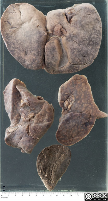
- The liver, lungs and spleen from a 3 month old child who died of extensive miliary tuberculosis.
- The miliary foci are a little larger than in the first example, but they seldom expand beyond a few mm since death intervenes before this can occur.


