Digital Pathology by University of Cape Town is licensed under a Creative Commons Attribution-NonCommercial-ShareAlike 4.0 International License. Permissions beyond the scope of this license may be available at www.pathologylearningcentre.uct.ac.za.
Case 1
Clinical data
- The patient was a 68 year old man who was admitted to hospital for cardiac failure.
- He developed acute urinary retention and had to be catheterised.
- He died of complications of vascular insufficiency.
Macroscopy pathology
- The bladder and prostate gland have been opened anteriorly.

- The median lobe of prostate is enlarged and nodular, and intrudes into the bladder floor.
- It obstructs the internal urethral opening, possibly with a ball-valve effect.

This patient’s bladder shows some of the secondary effects of urinary outflow obstruction.
- Trabeculations (ridging) are due to hypertrophy of the detrusor muscle. The inner surface of a normal bladder is quite smooth.
- Permanent distension. The normal adult bladder is elastic with a capacity of 300 - 500ml.

- Diverticuli are outpouchings of the bladder wall.
- At autopsy the patient was also found to have bilateral hydroureter and hydronephrosis (not shown).

Anatomical note
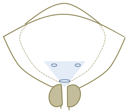
- The trigone of the bladder is a triangular region of the inner bladder defined by the ureteral orifices and the internal urethral orifice.

Case 2
Clinical data
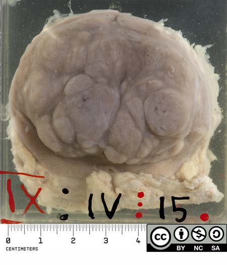
- This is the prostate of a 79 year old man
- He died of a ruptured aortic aneurysm and the abnormal prostate was an incidental finding at autopsy.
Macroscopic pathology
- The prostate is enlarged and coarsely nodular. (The normal prostate is the size of a walnut and weighs about 20g.)

This is a transverse section through the prostate.
- In this case it is predominantly the two lateral lobes that are hyperplastic

Terminology
- The bladder shows hypertrophy
-
The prostate shows hyperplasia
- What is the difference?
- What sort of organs tend to hypertrophy?
- What sort of organs lean to hyperplasia?
- Can you define hypoplasia or dysplasia or metaplasia, even aplasia?
- Similarly, dystrophy or atrophy?
- How would you explain to your patient with benign prostatic hyperplasia (BPH) what is happening in his prostate?
Histology
- The naked-eye appearance of benign prostatic hyperplasia was confirmed by microscopy.
- In addition, small foci of adenocarcinoma were seen.
Comment on this case:
- BPH is very common in men over 50 years.
- The finding of microscopic or latent prostatic carcinoma is very common in men over 70 years.
- The two conditions are independent i.e. BPH does not predispose to carcinoma.
- Benign hyperplasia involves both the glandular tissue and fibromuscular stroma of the prostate. It tends to occur in the central, peri-urethral region of the prostate.
- Prostate cancer is usually adenocarcinoma, deriving from glandular tissue. Most arise in the peripheral subcapsular region of the prostate, where the main glands are located.
References and links
- For a review of the diseases of the prostate and prostate histology, go to this excellent on-line tutorial http://library.med.utah.edu/WebPath/TUTORIAL/PROSTATE/PROSTATE.html

