2012
Nina Liebenberg
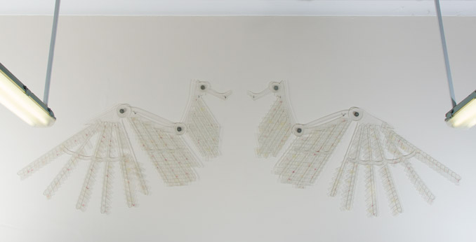
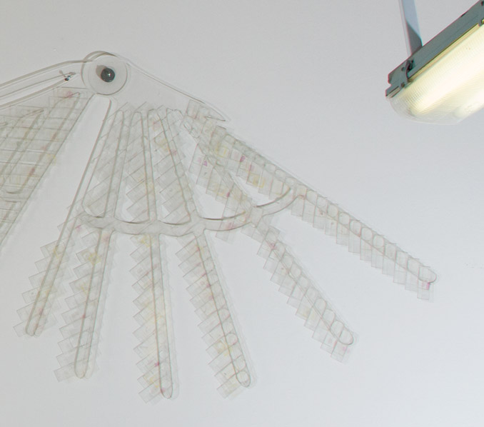
Histology slides, Perspex
Histology slides are prepared by taking a sample of biological tissue and fixing it to preserve the tissue in as natural a state as possible and prevent postmortem decay. The tissue is immersed in a chemical fixative and then embedded in wax to make it hard enough to cut into very thin sections of tissue (usually 5 to 7 micrometers in thickness). It is then passed through baths of solvents which remove the wax, then through graded alcohols, water and finally through baths of haematoxylin and eosin to stain it for better viewing under a microscope.

p>

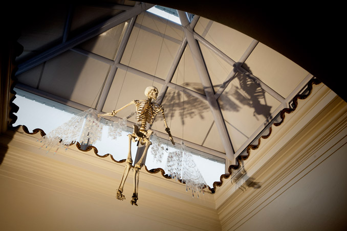
In the original conception of Icarus, these wings were attached to a skeleton. The skeleton had been sent to the Pathology Learning Centre with the Saint surgical pathology bottles. He had a broken stand and it was thought that he was a standard teaching skeleton possibly prepared in the anatomy workshop here at UCT or otherwise purchased. The Saint collection had been in a storeroom at the back of the surgery department for years. The storeroom had been closed all that time, however somebody had left a window open resulting in pigeons roosting in the room, their droppings covering everything, including the skeleton. In transit, many things got broken. They never found his hands.

