Poster Presentations at the EuroMedLab Conference in Brussels, Belgium - 2025
|
|
Registrar Dr KG Magolego |
Abstract:
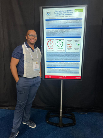
FROM LABORIOUS TO EFFORTLESS: TRANSFORMING AMINO ACID RESULT REPORTING WITH AUTOMATION
K.T.R. Magolego 1, J.P. Croxford 1, H. Vawda 1, B. Southon 1, J.A. Rusch 1
1Division of Chemical Pathology, Department of Pathology, University of Cape Town and National Health Laboratory Service, South Africa
BACKGROUND-AIM
The discontinuation of the amino acid kit at our laboratory necessitated outsourcing analysis to a referral laboratory. This laboratory provided amino acid results as PDF reports without interpretive comments. Following interpretation, results for 29 amino acids and an interpretive comment were manually uploaded to the Laboratory Information System (LIS) and verified by a second person to rule out transcription errors.
Manual result entry is a common challenge in laboratory medicine, arising from manual assays, results from external providers, or analysers lacking LIS interfacing. These processes are labour-intensive, time-consuming, and prone to various errors. To address these inefficiencies, we developed and verified an automated result entry and verification pipeline.
METHODS
The development of the Python-based script, supplemented with AutoHotkey, was an iterative process, with extensive troubleshooting to ensure it could handle the complexities of both Excel and LIS workflows seamlessly. The pipeline began with PDF-to-Excel data extraction and Z-score plotting, enabling rapid result visualisation to assist staff in generating interpretive comments. The script then integrates the results from the Excel files into the LIS. To ensure accuracy, it performs result verification by cross-referencing values extracted from the electronic patient record with the source dataset. During pipeline verification, all results were manually checked to confirm that no errors occurred.
RESULTS
Once developed, 503 serum and urine amino acid reports, including 29 analytes each and an interpretive comment, were used to verify the automation pipeline. Verification revealed zero errors. Result entry time decreased from 204 minutes to 53 minutes per batch, with batches consisting of 31 reports, on average. Verification time dropped from 85 minutes to 55 seconds.
CONCLUSIONS
We successfully automated the entry and verification of amino acid results, saving time, reducing workload, and eliminating transcription errors. This project demonstrates significant efficiency gains and provides a framework for other laboratories to adopt similar automation of tedious, manual tasks to improve workflows and reduce errors.
|
|
Registrar Dr Riona Singh-Gansen |
Abstract:
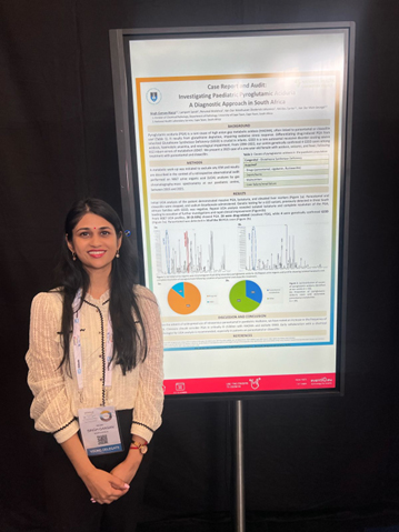
CASE REPORT AND AUDIT: INVESTIGATING PAEDIATRIC PYROGLUTAMIC ACIDURIA– A DIAGNOSTIC APPROACH IN
SOUTH AFRICA
R. Singh-Gansan 2, S. Lampert 3, M. Perumal 1, D.J. Van Der Westhuizen 2, S. Meldau 2, G. Van Der Watt 2
1Division of Chemical Pathology, Department of Pathology, National Health Laboratory Service, Cape Town, South Africa 2Division of Chemical Pathology, Department of Pathology, University of Cape Town and National Health Laboratory Service, Cape Town, South Africa
3Division of Chemical Pathology, Department of Pathology, University of Cape Town, Cape Town, South Africa
BACKGROUND-AIM
Pyroglutamic aciduria (PGA) is a rare cause of high anion gap metabolic acidosis (HAGMA), commonly associated with administration of paracetamol and/or cloxacillin derivatives. PGA impairs the cell’s ability to manage oxidative stress due to insufficient glutathione. Differentiation between drug induced causes and genetic disease such as Glutathione Synthetase Deficiency (GSSD) is critical in acute infantile presentations. The latter is an autosomal-recessive inborn error of glutathione metabolism, characterized by severe metabolic acidosis, haemolytic anaemia, and neurological complications. During 1994 – 2022, we have genetically confirmed 4 cases of GSSD from 4 families, among 812 inborn errors of metabolism at our centre.
We review a case from 2023,where a one-year-old female presented with acidosis, seizures, and pyrexia. She was treated with paracetamol and cloxacillin.
METHODS
A metabolic work-up was initiated to exclude an inborn error of metabolism (IEM) and results are described in the context of a retrospective observational audit performed on 9067 urine organic acid (UOA) analyses at our paediatric centre, between 2015 and 2021.
RESULTS
Initial UOA analysis of the patient demonstrated massive PGA, lactaturia, and elevated liver markers. Paracetamol and cloxacillin were stopped, and sodium bicarbonate administered. Genetic testing for a GSS variant, previously detected in 3 South African families with GSSD, was negative. Repeat UOA revealed isolated lactaturia and complete resolution of the PGA, leading to cessation of further investigations and rapid clinical improvement.
Results from our UOA analyses audit identified 30 out of 9067 profiles (0.33%) with PGA. Of these, 26 were attributed to drug use, prompting drug cessation, while 4 cases were investigated further. In 10 of the 30 positive UOA profiles, paracetamol was also present.
CONCLUSIONS
Since the advent of widespread use of intravenous paracetamol in paediatric medicine, we have noted an increase in the frequency of PGA. Clinicians should be aware of PGA as a cause of HAGMA in critically ill children and consider GSSD. They should have a low threshold to collaborate with a chemical pathologist to perform UOA analysis especially
in patients receiving paracetamol or cloxacillin derivatives.
AN AUDIT OF LIPOPROTEIN ELECTROPHORESIS WITH A FOCUS ON LIPOPROTEIN X: A CAPE TOWN PERSPECTIVE
R. Singh-Gansan 1, C. Francis 2, J. Pienaar 2, D.M. Blackhurst 2, A.D. Marais 2
1Division of Chemical Pathology, Department of Pathology, University of Cape Town and National Health Laboratory Service, Cape Town, South Africa
2Division of Chemical Pathology, Department of Pathology, University of Cape Town, Cape Town, South Africa
BACKGROUND-AIM
Lipoprotein X (LpX) is a pathological lipoprotein composed of phospholipids, non-esterified cholesterol and trace apolipoproteins but is deficient in neutral lipids and apolipoprotein B. It arises from cholestasis, lecithin–cholesterol acyltransferase deficiency or intravenous lipid infusions. Conjugated hyperbilirubinaemia and elevated hepatic canalicular enzymes accompany the hypercholesterolaemia. Complications include xanthomas, pseudohyponatraemia, and hyperviscosity syndrome. This study reviews our experience with LpX at a teaching hospital.
METHODS
Lipoprotein electrophoresis test results were extracted from the Laboratory Information System from 1 January 2016 to 19 November 2024 for a retrospective observational audit.
RESULTS
Among 1747 tests, Type 2a hyperlipidaemia was most frequent (740 cases), followed by Type 2b (426), normal profiles (220), Type 4 (182), Type 5 (87), Type 3 (62), Type 1 (4) and unclassified (17). LpX was identified in only 9 cases (8 adults and 1 child; 4 males and 5 females), highlighting its rarity. Total cholesterol, triglycerides, conjugated bilirubin, alkaline phosphatase, and gamma-glutamyl transferase results were elevated. One case had markedly increased bile acids. All cases had a low sodium result- likely due to pseudohyponatraemia. 3 cases had xanthomas. 7 cases noted a calculated/directly measured low-density lipoprotein (LDL) cholesterol result. One case, initially misclassified as Type 2a hyperlipidaemia, was correctly diagnosed with LpX by non-denaturing polyacrylamide gradient gel electrophoresis.
CONCLUSIONS
Laboratory diagnosis of LpX is challenging. Useful investigations include agarose electrophoresis showing no/ little migration from the application; non-denaturing gradient gel electrophoresis; measurement of non-esterified cholesterol relative to total cholesterol; and the cholesterol/apo B ratio. Unless LpX is excluded, LDL cholesterol should not be calculated in hypercholesterolaemia linked with cholestasis. Direct assays may also be unreliable. LpX interferes with several biochemical measurements. Very high cholesterol levels associated with conjugated hyperbilirubinaemia and canalicular enzyme induction should prompt clinicians to consider LpX and discriminate it
from other dyslipoproteinaemias.
SERUM POTASSIUM MEASUREMENT: DEPENDENCE ON SEASONAL TEMPERATURE AND INDEPENDENCE FROM PRE- CENTRIFUGATION DELAYS ≤ 30 HOURS
H. Vawda 1, D. Van Der Westhuizen 1
1Department of Chemical Pathology, NHLS, Groote Schuur Hospital and University of Cape Town, Cape Town
BACKGROUND-AIM
Hyperkalaemia is characterised by an elevation of serum potassium (K+) above 5.5 mmol/L. Pseudohyperkalaemia, a false elevation in K+, may result from pre-analytical errors, affecting clinical management. A common cause of pseudohyperkalaemia is delayed serum sample separation, with delays greater than 24 hours causing altered K+ results. Seasonal temperature changes may impact K+, but thorough investigation is limited. This study aims to analyse the effects of delay and seasonal temperature on K+ measurement.
METHODS
Serum K+ results from January to December 2023 were extracted from the laboratory information system, including 173023 patient samples from Groote Schuur and Red Cross War Memorial Children’s Hospitals’ National Health Laboratory Service (NHLS) laboratories in Cape Town, South Africa. Samples with a haemolysis >90 mg/dL and those lacking a collection time were excluded. The time between sample collection and registration in the lab was calculated as the pre-centrifugation delay. Cape Town monthly temperatures were retrieved from the World Weather website.
RESULTS
Data analysis showed similar average centrifugation delays across all months. The average K+ levels in the warmer months (November to April) had statistically significant differences compared to cooler months (June to September), p<0.05. There was a significant negative linear relationship between temperature and K+ (R2=0.909, p<0.0001). The relationship between time and K+ showed weak linear regression (R2=0.1733, p=0.178). Of the 401 samples with a centrifugation delay ≥30 hours, only 76 (19%) had clinically significant K+ values.
CONCLUSIONS
Serum K+ samples remain stable for up to 30 hours without centrifugation. Seasonal temperature variations have a greater impact on K+levels than centrifugation delays. Clinicians and pathologists in South Africa should account for these seasonal fluctuations when interpreting K+ results. The NHLS may need to reassess their rejection policy for serum K+ samples between 24 and 30 hours, and take into account the influence of temperature fluctuations on K+measurement.