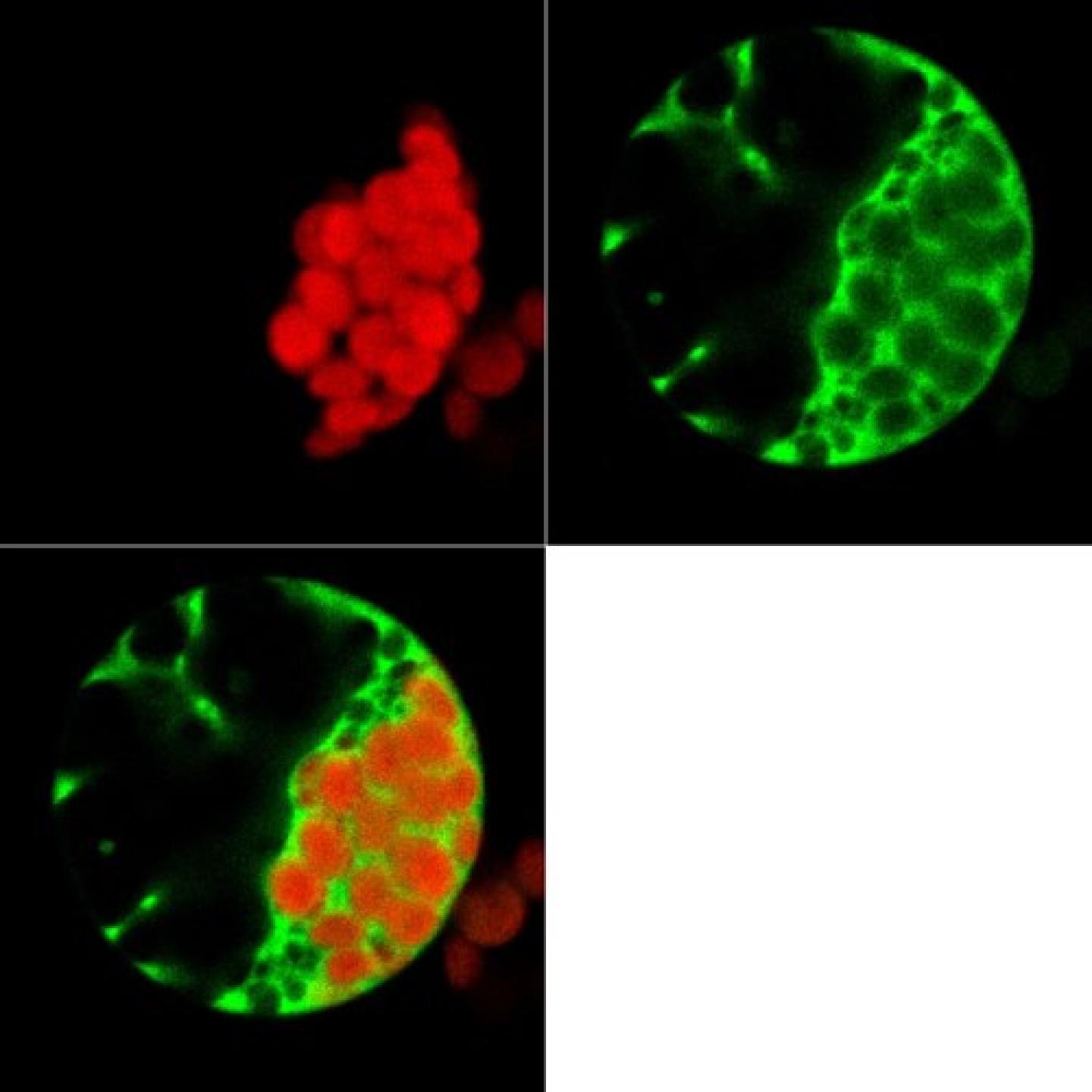
Expression of a GFP-tagged cytoplasmic protein (green) in a plant protoplast. Chloroplasts in red
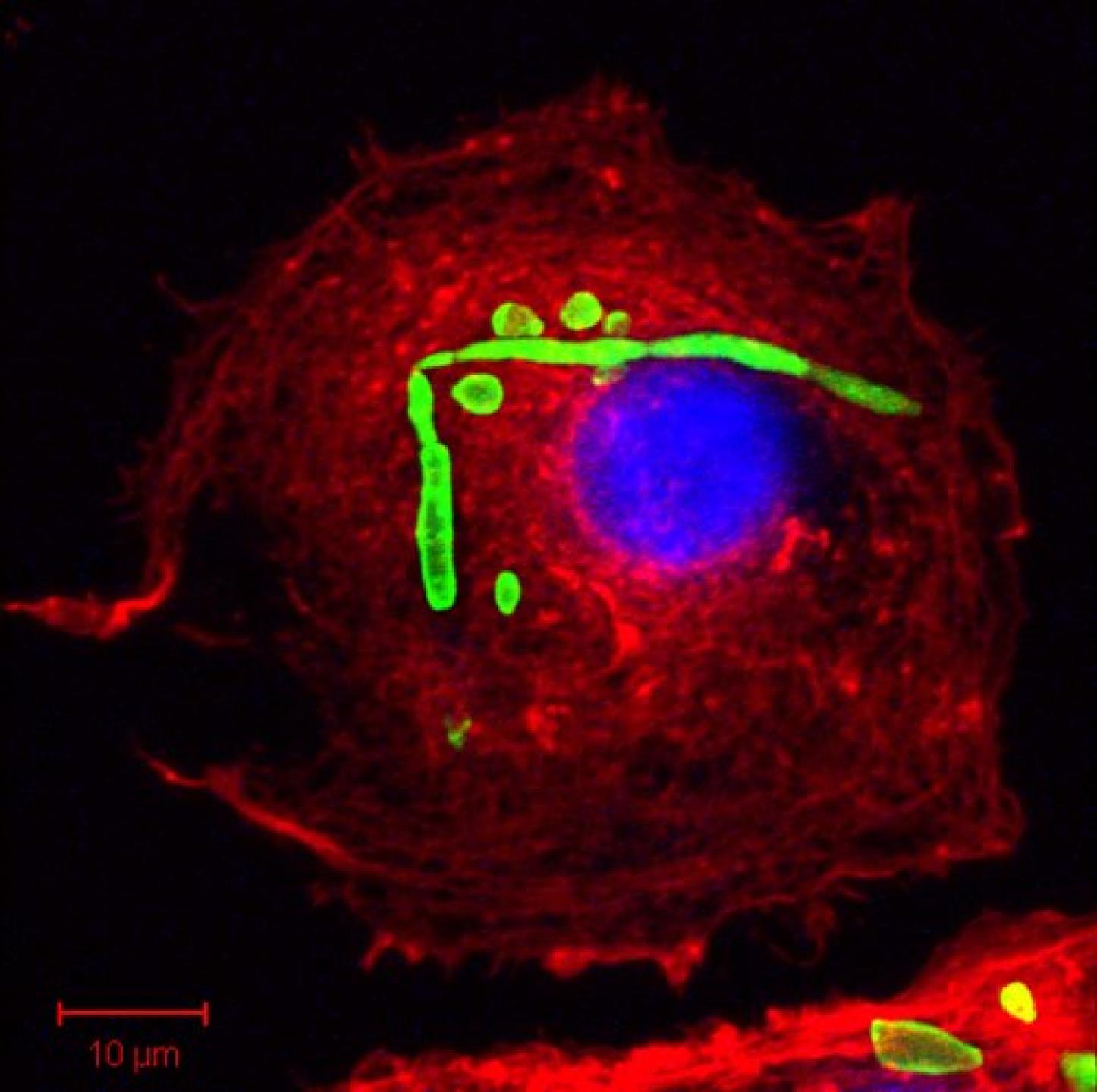
Phalloidin-labelled (red) macrophage showing internalized fungus (GFP, green). DAPI nuclear stain (blue).
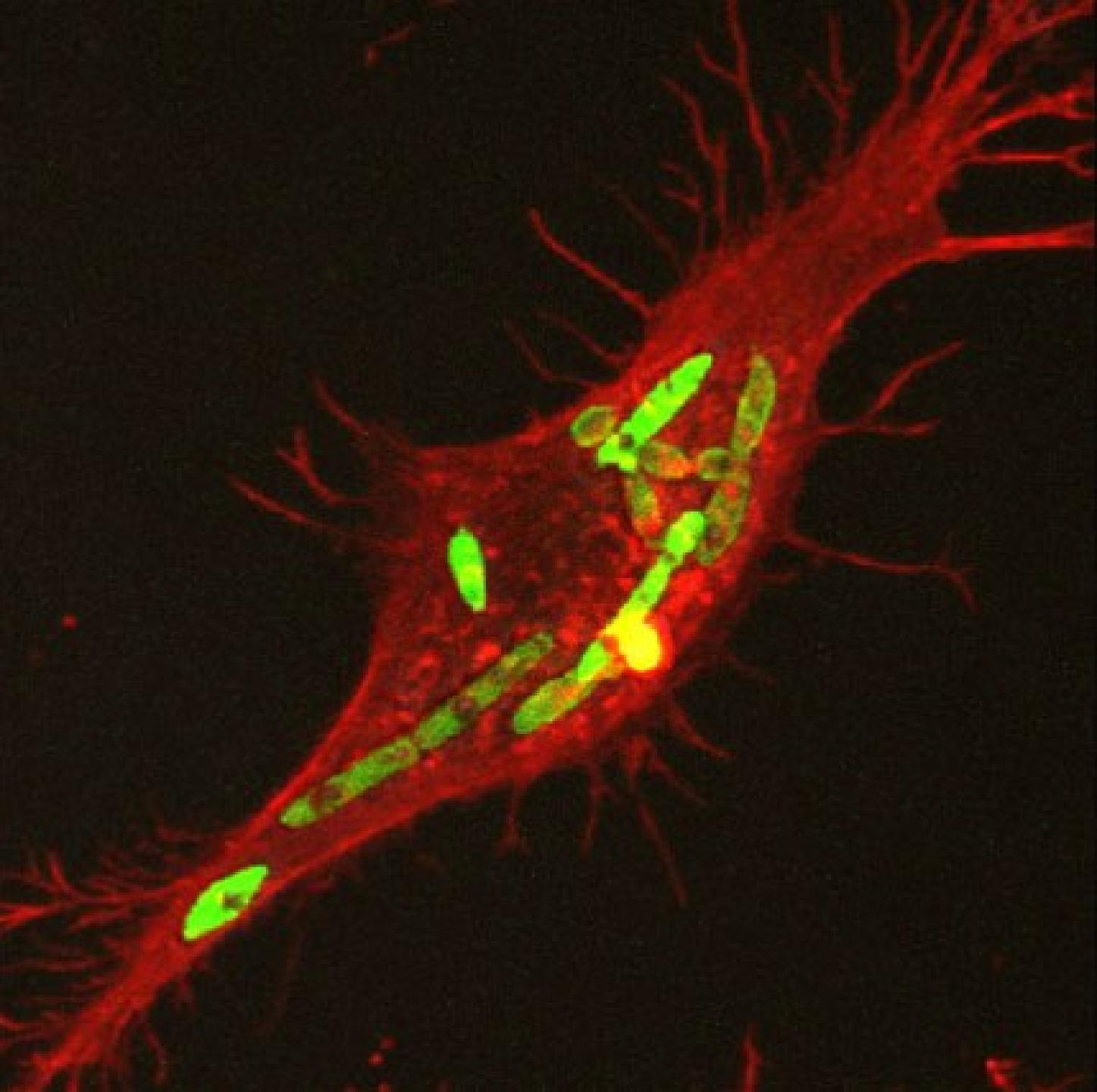
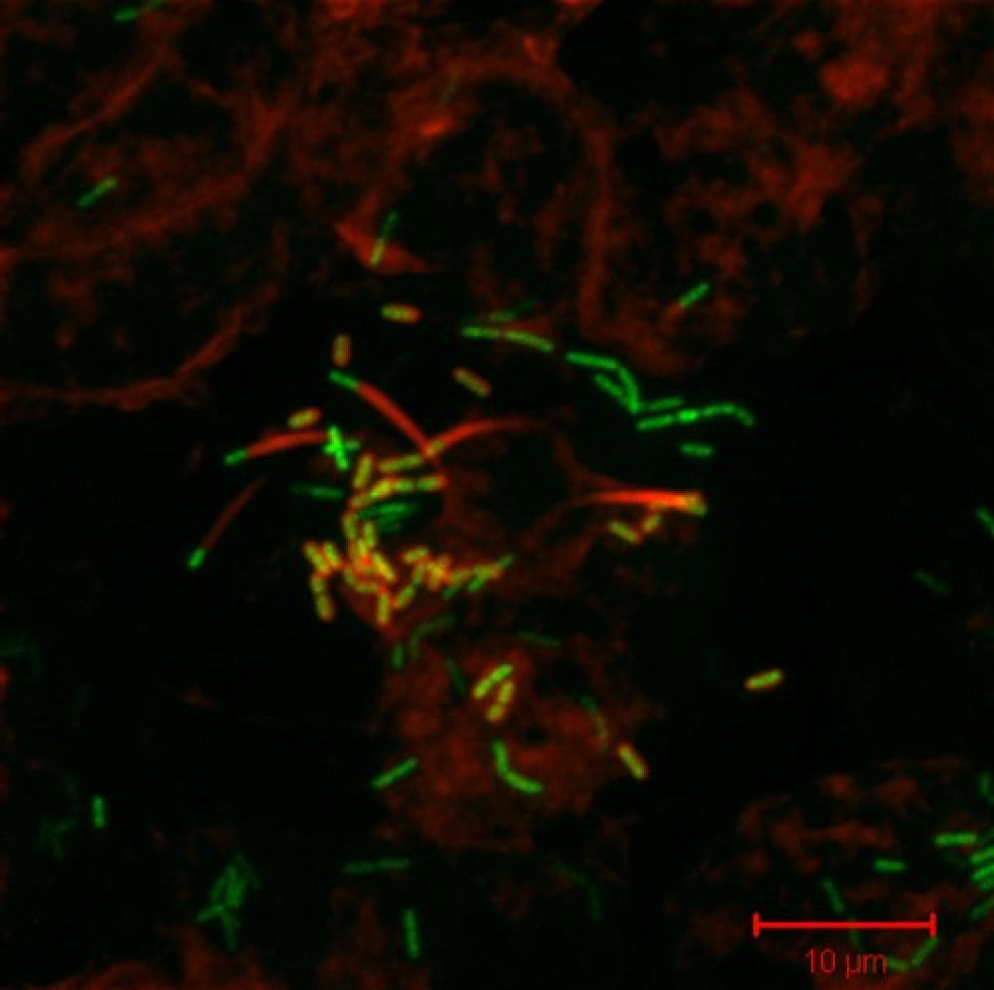
Phalloidin-labelled (red) macrophage containing fungal hyphae (green)
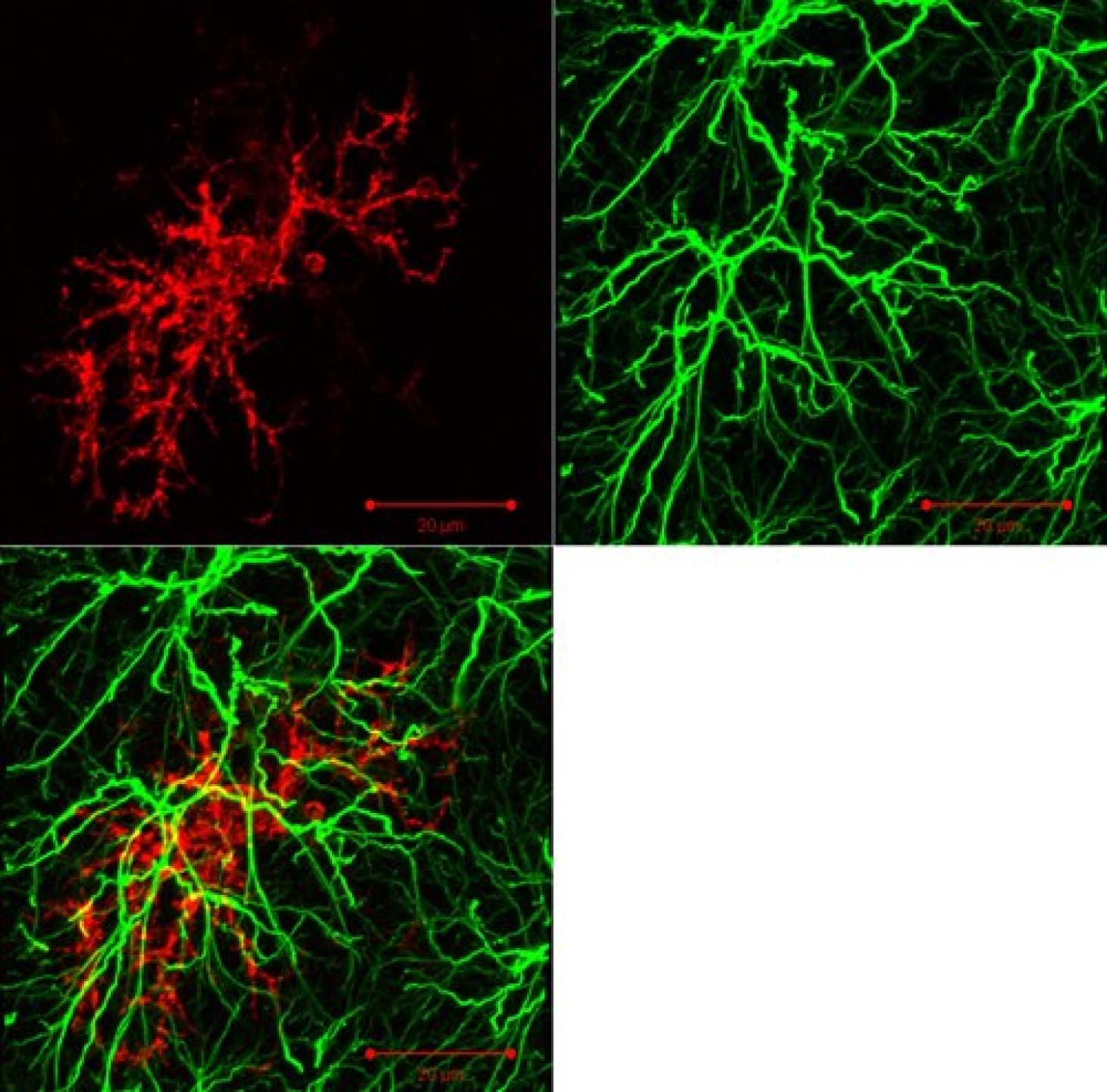
Immunostaining of microglia (red) and astrocyte processes (green) in a section of rat brain.
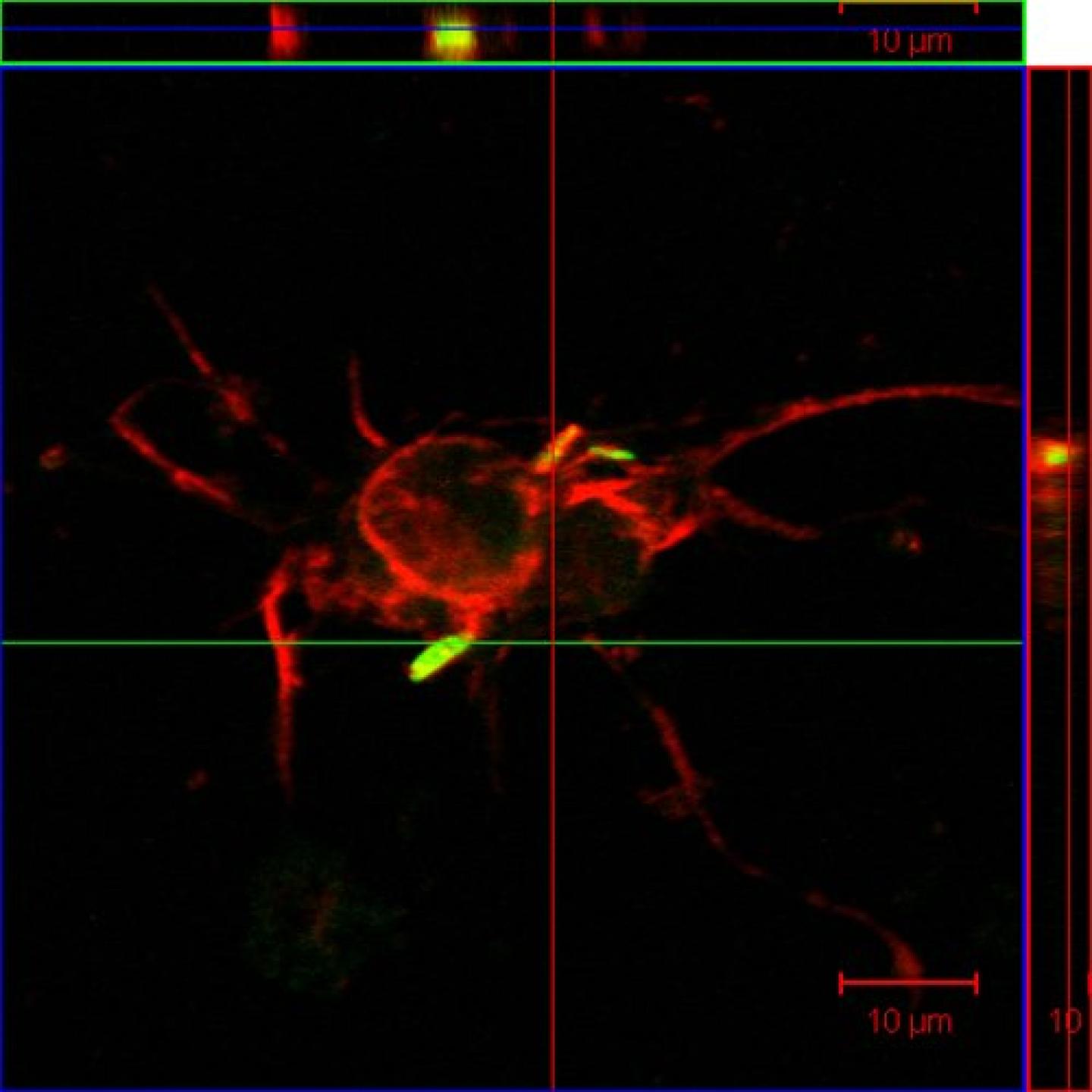
Neurons (labelled with anti-MAP-2 antibodies, red) internalize GFP-tagged TB bacilli (green), orthogonal projection.
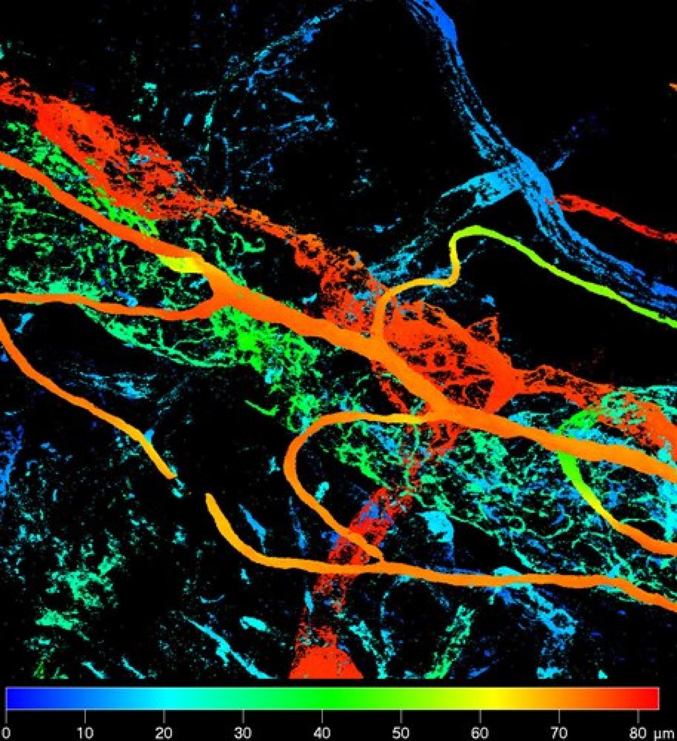
Depth-coded false colour projection of immunolabelled blood vessels in the anterior segment of the eye.
GFP-tagged Listeria (green) form actin "comets" (phalloidin-rhodamine labelled, red) in infected macrophages.
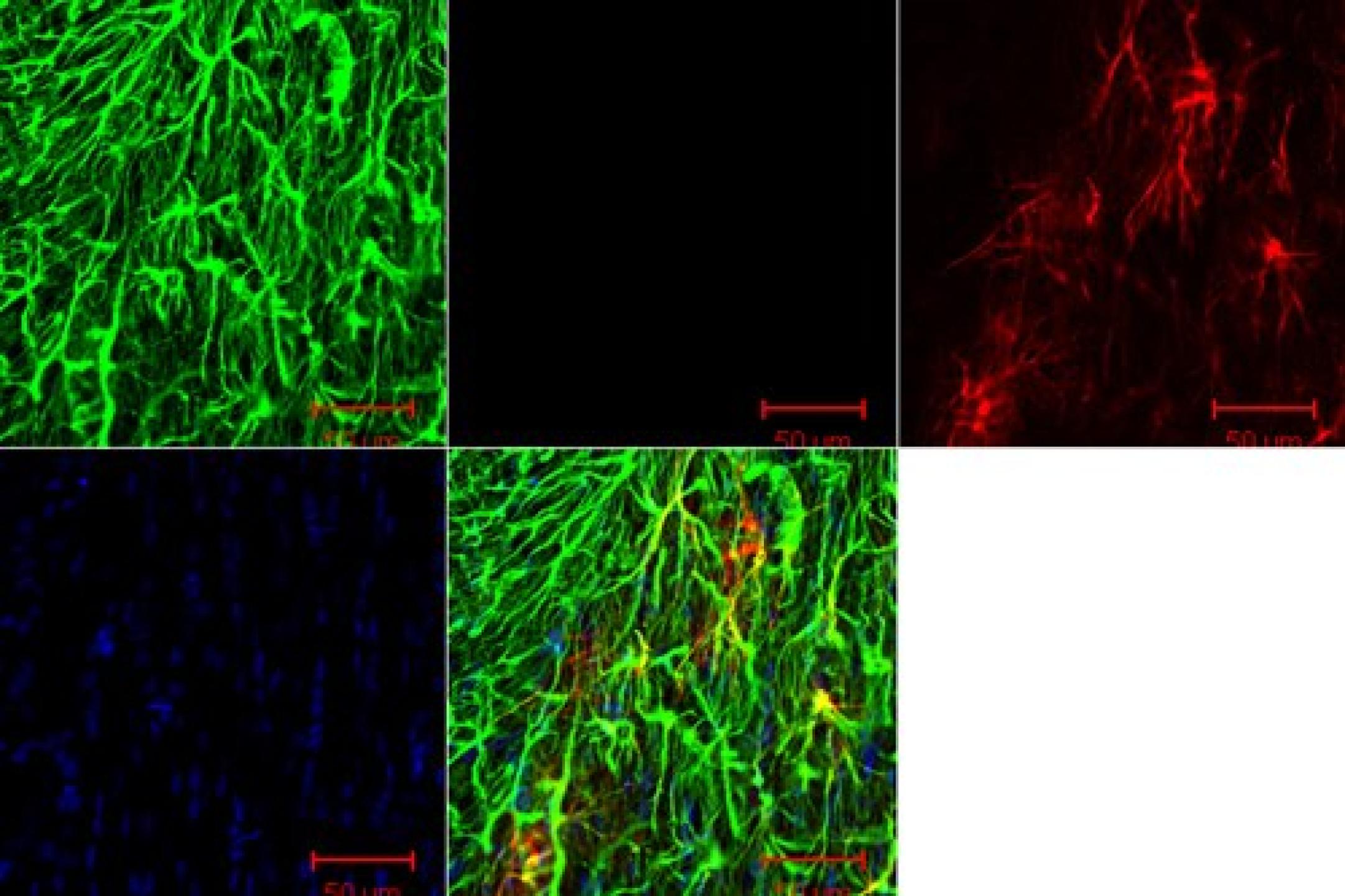
Astrocytes in a rat brain section, identified by anti-GFAP antibody labelling (green), show Heat Shock Protein (HSP) 25 immunoreactivity (red) after neurotoxic insult with 6-OHDA. Hoechst nuclear label (blue).
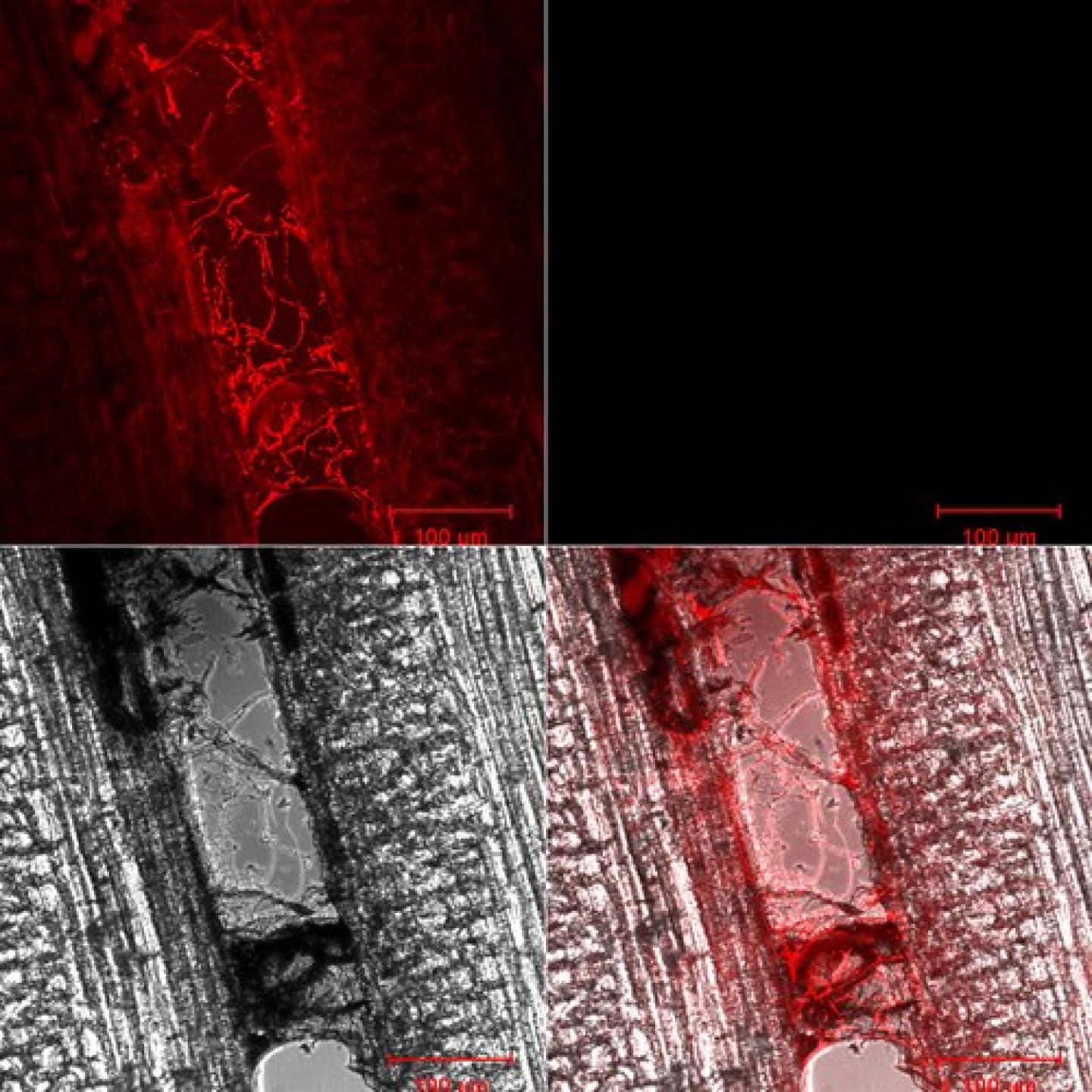
A vine (DIC) showing infection with a DS-Red-tagged fungus.
Expression of a GFP-tagged cytoplasmic protein (green) in a plant protoplast. Chloroplasts in red
Phalloidin-labelled (red) macrophage showing internalized fungus (GFP, green). DAPI nuclear stain (blue).
Phalloidin-labelled (red) macrophage containing fungal hyphae (green)
Immunostaining of microglia (red) and astrocyte processes (green) in a section of rat brain.
Neurons (labelled with anti-MAP-2 antibodies, red) internalize GFP-tagged TB bacilli (green), orthogonal projection.
Depth-coded false colour projection of immunolabelled blood vessels in the anterior segment of the eye.
GFP-tagged Listeria (green) form actin "comets" (phalloidin-rhodamine labelled, red) in infected macrophages.
Astrocytes in a rat brain section, identified by anti-GFAP antibody labelling (green), show Heat Shock Protein (HSP) 25 immunoreactivity (red) after neurotoxic insult with 6-OHDA. Hoechst nuclear label (blue).
A vine (DIC) showing infection with a DS-Red-tagged fungus.