Members of the family herpesviridae are found in a wide range of host systems. To date, at least seven different species are known to infect man, including herpes simplex virus (HSV); cytomegalovirus (CMV), varicella zoster (VZV); and Epstein Barr virus (EBV).
Herpesviruses have an
envelope surrounding an icosahedral
capsid, approximately 100nm in diameter, which contains the dsDNA genome.
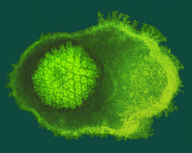
When the envelope breaks and collapses away from the capsid, negatively stained virions have a typical "fried-egg" appearance.
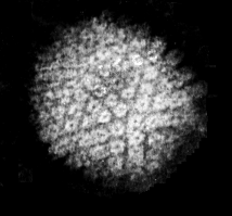
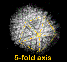
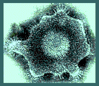
The herpes virus CAPSID is an icosahedron of triangulation number T = 16. There are 12 pentavalent capsomers (one at each apex) and 150 hexavalent capsomers. Each capsomer has a deep central indentation.
The
ENVELOPE that surrounds the nucleocapsid is derived from the inner nuclear membrane of the host cell.Virus-encoded glycoproteins are incorporated into the virion envelope and are visible as "spikes" that project from its surface.
In the micrograph of HSV, the glycoprotein B (gB) is clearly visualised in clusters of spikes appoximately 10 nm in length.
Between the capsid and the envelope is an ill-defined layer of proteins, collectively known as the tegument.
© Copyright Dr Linda M Stannard, 1995
This page was written by Dr Linda Stannard, on behalf of the Division of Medical Virology, UCT.
In Memory of Dr Linda Stannard, 10 May 1942 - 17 October 2016
For re-use of and queries about Dr Stannard's images, please contact Dr Stephen Korsman at stephen.korsman@uct.ac.za.
Recipient of Key Resource Award


